2 years ago Maxillofacial surgery
How to diagnose and treat fractures of the zygomatic-orbital complex and zygomatic arch (bone)?

However, powerful blows to the face that cause a fracture of the maxillo-orbital complex can also cause brain trauma. Therefore, before the maxillofacial surgeon begins the operation, the patient must be examined and examined by a neurosurgeon, as well as an anesthesiologist. In addition, the patient passes preoperative tests on an empty stomach. All this takes time, and if the operation was not done within the first day, it is possible after five or six days, when the post-traumatic swelling subsides.
What is a zygomatic fracture?
A fracture of the zygomatic bone occurs when the face is hit from the side (it is not necessarily a boxing blow, it can also be an unfortunate fall, a road accident or a domestic injury). At the same time, the process of the cheekbone, which goes from the cheekbone to the ear, breaks. With such a fracture, there are not so many fragments, so it is easier to heal.
Causes of a fracture of the zygomatic-orbital complex
Fracture of the maxillo-orbital complex is a complex injury. When all three processes of the zygomatic bone are broken and displaced, we get a fracture of the outer wall of the orbit, its lower edge, the front wall of the maxillary sinus, the zygomatic alveolar ridge, and the zygomatic arch. If the zygomatic bone separates from the skull, it is always a complex comminuted fracture with complications. The cause of such a displaced fracture of the zygomatic bone is a blow to the face from the front (in a road accident, combat or household injury, fall).
What are the signs of a fracture of the zygomatic arch?
If you receive any trauma to the facial bones, you should immediately consult a maxillofacial surgeon. Signs of a zygomatic arch fracture are:
- numbness of a part of the face after a blow;
- bleeding from the nose (if the brain is also affected, blood can flow together with cerebrospinal fluid);
- swelling under the eye;
- pain when trying to open the mouth;
- facial asymmetry.
During the first one to two hours after the injury, the side where the facial bone is broken appears sunken inward. However, as edema develops, it swells and becomes larger than healthy.
Types of fractures of the zygomatic-orbital complex
Fractures of the zygomatic-orbital complex are classified according to a number of signs. In particular, the following types of fractures of the zygomatic-orbital complex and the zygomatic bone are distinguished:
- according to the presence of an external wound: closed and open;
- according to the degree of severity of the fracture of the zygomatic bone: single and multiple;
- according to the presence of displacement: fracture of the zygomatic bone without displacement or with displacement;
- by the number of fragments: fragmentary and linear.
Fractures of the facial bones are also distinguished by the place of injury: fractures of the jaws, zygomatic-orbital complex, lower edge of the orbit, bridge of the nose.
How is a cheekbone fracture diagnosed?
The best method for diagnosing a fracture of the maxillo-orbital complex is computed tomography. The fracture of the zygomatic bone and the position of the fragments are clearly visible on the CT scan, the surgeon can determine the optimal access and scope of the future operation. Since such a fracture is often accompanied by craniocerebral trauma (concussion, hemorrhage), it is necessary to describe not only the facial skull, but also the brain, so CT scan of the head will be optimal.
In a certain projection, the X-ray also shows the presence of a fracture of the zygomatic bone. But the fractures are usually combined, and in addition to the zygomatic bone, the lower edge of the orbit, the zygomatic alveolar ridge, the front and lateral walls of the maxillary sinus may be broken, which is not visible on X-ray. So these pathologies risk remaining undiagnosed and, as a result, incurable.
Methods of treatment of facial arch fracture
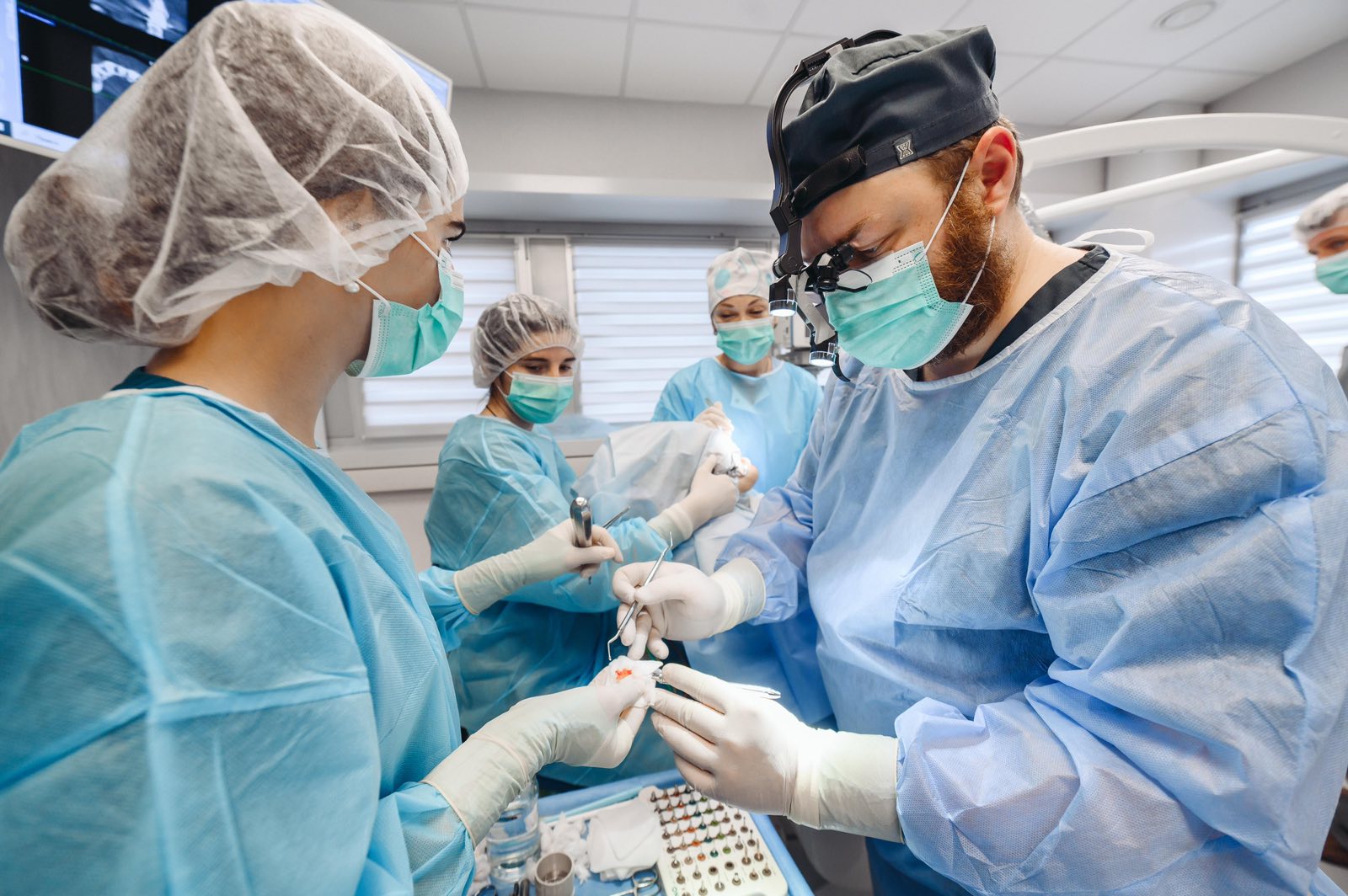
Treatment of a fracture of the zygomatic bone in the Sirius Dent Medical Center is carried out by the method of metal osteosynthesis. If during a bone fracture, the fragments pinched the vascular-nerve bundle innervating the infraorbital region, an important task of the surgeon is to release it, so that the patient does not get permanent paresthesia of the face. The maxillofacial surgeon must place the bone fragments in the correct places and secure them with titanium plates and screws. In the case of an open wound and the loss of part of the fragments, they are reproduced in a special computer program and printed from titanium on a 3D printer.
The task of the maxillofacial surgeon is to restore the person’s pre-injury appearance as much as possible without leaving scars, and if this is possible, repositioning of most fragments is done through an intraoral approach. In some cases, an incision is made along the lower edge of the eyelid or elsewhere, depending on where it is more convenient to reach the bone fragments. Surgery for a fracture of the zygomatic bone lasts one to three hours, after which the patient remains under medical supervision in the dent medical center for two to three days and returns home.
Complication of damage to the zygomatic-orbital complex
Complications can vary depending on the nature of the fracture. In the case of a fracture of the lower wall of the orbit, where the thickness of the bone is no more than one millimeter, the fragments fall into the maxillary sinus, and the eye “slides” there – as a result, the patient will have permanent visual impairment (enophthalmos). And if blood and bone fragments are not removed from the maxillary sinus in time, chronic inflammation and suppuration of the hematoma are formed.
In cases where fragments of a broken bone have pinched or damaged the inguinal nerve, this leads to loss of sensitivity of the face and upper jaw. In general, treatment not carried out (an old fracture of the zygomatic bone) or performed poorly (improperly fused fracture of the zygomatic bone) leads to noticeable asymmetry of the face.
What are the limitations after treatment of a zygomatic fracture?
After treatment of the cheek bone, you should protect your face and avoid physical exertion. One week after surgery, the maxillofacial surgeon removes the stitches, and three months later, he orders a follow-up CT scan to check how the bone has grown. After that, it is possible to remove the titanium plates, which are placed close to the surface of the skin and are felt to the touch. However, these plates usually do not cause any discomfort and do not even react to metal detectors.
popular questions:
How much does a fracture of the zygomatic bone grow?
The zygomatic fracture heals within three weeks, but in addition, the bone tissue must regain density and strength. This process lasts about half a year.
Is it possible to do boxing after a fracture of the cheekbone?
Boxing and other contact sports can be practiced six months after a fracture of the zygomatic bone. Before that, it is necessary to do a control CT scan to make sure that the repositioning of the fragments went well and the bones fused correctly.
Share this content:

The author of the article: Valkevich Denis
Head of the Sirius Dent Medical Center, maxillofacial surgeon, orthopedic dentist, the implant surgeon
Similar articles

1 year ago
How to treat a facial bone fracture?
1 year ago
How is surgical treatment of a jaw fracture carried out?
2 years ago
Types and methods of treatment of neoplasms of the parotid salivary glandsother topics
Ask a Sirius-Dent expert at the consultation

